Breast Seed Brachytherapy
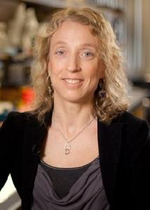
Juanita Crook MD FRCPC, Radiation Oncologist, BC Cancer Agency Sindi Aluwalia Hawkins Centre for the Southern Interior
| Total Number of Gifts: 7 | |
| Total Value of Gifts: $1,120.00 |
- Tell a friend about
- Create a page like this
- Please bookmark this page.
Recent Donors
Gail & Ian Blackford
Anonymous
Jim & Joanne Ross
Anonymous
Mrs. Sue Hughes
Janice Schweizer
Brian Culbert
Support vital research
Breast seed brachytherapy delivers radiation treatment precisely to the region of the breast where the cancer was removed, minimizing radiation exposure to the rest of the breast and surrounding structures.
The treatment can be delivered in a single one to two hour out-patient procedure with the same effectiveness of standard radiation, which takes three and a half to five weeks. The procedure involves implanting radioactive seeds smaller than a grain of rice around the cancer site. The dose of radiation is then gradually released over a two-month period, preserving the surrounding healthy tissue.
In 2013, radiation oncologist Dr. Juanita Crook commenced a pilot program to determine the proportion of patients eligible for breast seed brachytherapy and what proportion of eligible women would choose this type of treatment instead of standard whole breast radiotherapy and HDR breast brachytherapy.
- To date, 50 patients have undergone the procedure;
Results from this cohort are being compared to women receiving high-dose rate (HDR) brachytherapy, with emphasis on patient satisfaction, quality of life and cosmesis. To date, 94% of respondents with more than 6 months follow up are very satisfied or totally satisfied with their treatment and fewer than 5% of those women suitable for this treatment elect to have standard external beam treatment instead.
- This initiative has been expanded to include an additional 50 patients.
- The team is currently examining post-procedure CT scans to calculate seed stability and dose distributions in comparison to HDR brachytherapy;
- Dr. Crook and her team are continuing to refine and adapt the technique for different breast sizes and tumour locations and incorporate new technology with 3D ultrasound in collaboration with colleagues from the University of Western Ontario.
If you think this page contains objectionable content, please inform the system administrator.